



How Vets Diagnose FCE in Dogs
One of the first questions every family asks is: “How do the vets know it’s FCE?”
A note about our site:
Where possible, FCEtalk uses minimal images for easier printing
helping you create clear, practical reference guides to support your pet’s recovery journey.

How Vets Diagnose FCE (Fibrocartilaginous Embolism) in Dogs
Understanding the tests, examinations, and process your veterinarian uses to diagnose a canine spinal stroke.
One of the first questions every family asks is:
“How do the vets know it’s FCE?”
When your dog suddenly loses the ability to walk, drags a limb, or collapses without warning, it can feel like your world has stopped. The answer is reassuringly straightforward—but not always immediate.
Unlike a broken bone that appears on an X-ray or an infection confirmed with a single blood test, Fibrocartilaginous Embolism (FCE) is diagnosed by carefully gathering evidence while ruling out other conditions that can cause similar symptoms.
Your veterinarian combines your dog’s history, a detailed neurological examination, diagnostic imaging such as MRI, laboratory tests, and ongoing observation to build the clearest possible picture. Although waiting for answers can be difficult, this systematic approach helps ensure your dog receives the most appropriate treatment and rehabilitation plan.
The encouraging news is that many dogs with FCE go on to make significant improvements with supportive care and rehabilitation.
Why Diagnosing FCE Can Be Challenging
One of the most confusing parts of the FCE journey is that there isn’t a single test that simply says:
“Yes, your dog has FCE.”
Instead, veterinarians diagnose FCE through a combination of:
- Your dog’s history
- Clinical signs
- Neurological examination
- Diagnostic imaging
- Excluding other diseases
This is known as a diagnosis by exclusion. That doesn’t mean your veterinarian is guessing. It means they’re using the same careful medical process that specialists use for many neurological conditions in both veterinary and human medicine.
Why Getting the Correct Diagnosis Matters
Several spinal conditions can initially look very similar.
These include:
- Intervertebral Disc Disease (IVDD)
- Spinal fractures
- Spinal tumours
- Infections
- Inflammatory diseases
- Degenerative conditions
- Bleeding around the spinal cord
- Rare vascular disorders
Although these conditions may all cause sudden weakness or paralysis, their treatments can be very different. Obtaining the most accurate diagnosis possible helps your veterinary team recommend the safest treatment plan for your dog.
For example:
- Some require surgery.
- Some require medications.
- Others require long-term medical management.
- FCE is generally managed with supportive care and rehabilitation rather than surgery.
Neurological Examination
The neurological examination is often the single most important step in diagnosing FCE. Unlike blood tests or scans, this examination provides immediate information about where the spinal cord has been affected and how severely it has been injured. Although it may look simple from the outside, every movement your veterinarian observes is providing valuable clues.
Watching Your Dog Move
Before touching your dog, your veterinarian will often spend time simply watching.
Observations help identify which limbs are affected and whether the problem appears to originate in the brain, spinal cord, nerves or muscles. They may observe:
- Standing posture
- Walking pattern
- Balance
- Coordination
- Foot placement
- Ability to bear weight
- Tail movement
Testing Reflexes
Next comes a series of neurological reflex tests. Each reflex provides information about specific spinal cord segments.
These may include:
- Patellar (knee) reflex
- Withdrawal reflex
- Muscle tone
- Tail reflexes
- Perineal reflex
- Limb positioning
Assessing Conscious Proprioception
One commonly performed test involves gently turning your dog’s paw so the top rests on the ground. A healthy dog quickly corrects the paw position. Dogs with spinal cord injury may take longer—or be unable—to reposition it. This helps determine how well nerve pathways are functioning.
Checking Pain Perception
One of the most important parts of the examination is evaluating sensation. This assessment provides valuable information about the severity of spinal cord injury and helps guide prognosis. These tests are performed carefully and only by trained professionals.
Your veterinarian may test whether your dog can:
- Feel light touch
- Feel deeper pressure
- Respond appropriately to painful stimuli
Determining the Location of the Injury
One goal of the neurological examination is called neuro-localisation. Rather than simply identifying that something is wrong, your veterinarian works out approximately where in the spinal cord the problem is located. Different spinal cord regions produce different patterns of weakness and reflex changes. Knowing the likely location helps determine:
- Whether MRI is needed
- Which spinal diseases are most likely
- Whether surgery might be appropriate
- What recovery expectations may be
Your Dog’s History
The history you provide is incredibly valuable. In many cases, your observations before arriving at the veterinary clinic become one of the strongest pieces of evidence supporting an FCE diagnosis. Try not to worry if you cannot remember every detail perfectly. Even a general description of what happened can be very helpful.
Your veterinarian may ask questions such as:
- When did the symptoms start?
- Was your dog running or playing?
- Did the collapse happen suddenly?
- Has anything like this happened before?
- Was your dog crying in pain?
- Did symptoms worsen after onset?
- Has your dog been eating normally?
- Has there been any recent trauma?
- Have toxins been possible?
- Has bladder function changed?
Sudden Onset Is an Important Clue
One feature that often points toward FCE is the sudden onset of symptoms. Although activity itself does not cause FCE, many episodes appear immediately after exercise or excitement.
Many owners describe events such as:
“She jumped off the couch.”
“He was chasing the ball.”
“Everything was normal five minutes earlier.”
Pain That Improves Quickly
Many dogs briefly vocalise when the spinal injury first occurs. However, unlike several other spinal conditions, ongoing severe pain is often absent after those first moments. This pattern helps veterinarians distinguish FCE from diseases such as IVDD, although every dog is different.
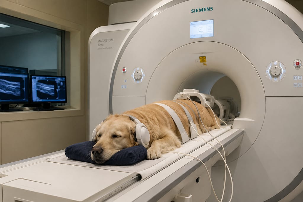
MRI
An MRI (Magnetic Resonance Imaging) is considered the best imaging test for evaluating the spinal cord.
While not every dog requires an MRI, it is often recommended when the diagnosis is uncertain or when surgery is being considered.
Why MRI Is So Helpful
Unlike X-rays, MRI shows the soft tissues inside the spinal canal, including:
- Spinal cord
- Intervertebral discs
- Nerves
- Inflammation
- Swelling
- Compression
- Bleeding
This level of detail helps veterinarians determine what is affecting your dog’s spinal cord.
What MRI Can Show in FCE
Dogs with FCE often have characteristic changes within the spinal cord itself. Importantly, MRI findings must always be interpreted alongside your dog’s examination and history.
MRI may reveal:
- Areas of spinal cord swelling
- Changes consistent with spinal cord infarction
- Localised injury
- No evidence of disc compression
- No spinal fracture
MRI Helps Rule Out Other Diseases
Sometimes MRI is just as valuable for what it doesn’t show. This helps veterinarians become more confident that FCE is the most likely diagnosis. It can exclude conditions such as:
- Disc extrusion
- Spinal tumours
- Vertebral fractures
- Compression requiring surgery
- Some infections
Does Every Dog Need an MRI?
Not necessarily.
Some dogs are diagnosed based on:
- Typical history
- Neurological findings
- Stable clinical progression
- Absence of ongoing pain
However, referral to a neurologist and advanced imaging may still be recommended if there is uncertainty or if your dog is not progressing as expected. Your veterinarian will discuss whether MRI is appropriate based on your dog’s individual circumstances.
Blood Tests
Many families are surprised when blood tests come back normal. This is actually quite common with FCE. That’s because blood tests generally do not diagnose FCE directly. Instead, they help evaluate your dog’s overall health and rule out other possible causes of neurological disease.
What Blood Tests Can Detect
Blood work may identify:
- Infection
- Inflammation
- Organ disease
- Electrolyte abnormalities
- Metabolic disorders
- Tick-borne diseases (where relevant)
- Underlying medical conditions affecting treatment
This information helps veterinarians plan anaesthesia, medications, hospitalisation and rehabilitation safely.
Why Normal Blood Tests Can Be Good News
Normal results often support the idea that another systemic illness is not responsible for your dog’s neurological signs.
Although they cannot confirm FCE, they provide important reassurance that many other diseases are less likely.
Diagnosis by Exclusion
One phrase families often hear is:
“We think this is FCE because we’ve ruled out the other possibilities.”
This can initially sound frustrating.
In reality, diagnosis by exclusion is a respected and widely used medical approach.
What Does Diagnosis by Exclusion Mean?
It means your veterinarian carefully considers every reasonable possibility before arriving at the diagnosis that best matches all available evidence.
This involves combining:
- History
- Neurological examination
- MRI findings
- Blood tests
- Progress over time
- Response to supportive care
The more pieces that fit together, the more confident your veterinary team becomes.
Why Time Can Help Confirm the Diagnosis
One interesting feature of FCE is that dogs often stabilise after the initial event.
Unlike some diseases that progressively worsen over days, many dogs with FCE stop deteriorating after the spinal injury occurs.
Over subsequent days and weeks, gradual improvement may begin.
That recovery pattern provides another important clue supporting the diagnosis.
Specialist Referral
If uncertainty remains, your veterinarian may recommend referral to a veterinary neurologist. Neurologists have additional training in diagnosing diseases affecting the brain, spinal cord and nerves. Seeking specialist advice does not necessarily mean your dog’s condition is worse—it simply provides access to additional expertise and diagnostic tools.
Referral may include:
- Advanced neurological examination
- MRI
- Cerebrospinal fluid (CSF) analysis if appropriate
- Ongoing rehabilitation recommendations
What Happens After Diagnosis?
Once FCE becomes the most likely diagnosis, the focus shifts from finding the cause to supporting recovery. Recovery is rarely a straight line. Some days will feel encouraging. Others may feel frustrating. That is completely normal.
Many dogs continue making progress for weeks or even months after their spinal injury.
Treatment commonly includes:
- Nursing care
- Assisted standing
- Physiotherapy
- Hydrotherapy when appropriate
- Bladder management
- Preventing pressure sores
- Nutrition
- Home modifications
- Regular reassessment
Supporting Your Dog While Waiting for Answers
Waiting for a diagnosis can be emotionally exhausting. It’s natural to search for certainty, especially when your dog’s future feels uncertain. Remember that an accurate diagnosis is worth taking the time to establish. It helps ensure your dog receives care tailored to their individual needs.
While your veterinary team completes the diagnostic process, you can help by:
- Keeping notes about changes in mobility
- Recording bladder and bowel function
- Following nursing instructions carefully
- Avoiding unapproved exercises
- Asking questions whenever you’re unsure
- Focusing on your dog’s comfort and wellbeing
Frequently Asked Questions
Can FCE be diagnosed without an MRI?
Sometimes. If your dog’s history, neurological examination and clinical progression are highly consistent with FCE, your veterinarian may make a presumptive diagnosis. However, MRI remains the best imaging test for confirming spinal cord changes and ruling out other conditions.
Why didn’t my dog’s blood tests show FCE?
Because FCE affects the spinal cord rather than causing changes that are typically detectable in routine blood work. Blood tests are primarily used to assess your dog’s overall health and exclude other illnesses.
Can X-rays diagnose FCE?
No. Standard X-rays are useful for assessing bones but cannot show the spinal cord itself. They may help identify fractures or other skeletal abnormalities but cannot confirm FCE.
Why is surgery usually not recommended for FCE?
Unlike some spinal conditions, FCE is not typically caused by ongoing compression of the spinal cord that can be relieved surgically. Treatment usually focuses on supportive care, rehabilitation and allowing the spinal cord time to recover.
Should I see a veterinary neurologist?
Many dogs benefit from referral, particularly if the diagnosis is uncertain, advanced imaging is needed, or your veterinarian recommends specialist input. Your primary veterinarian can help determine whether referral is appropriate.
Can the diagnosis change later?
Occasionally. If new symptoms develop or your dog’s recovery does not follow the expected pattern, your veterinarian may recommend further investigation to reassess the diagnosis.
Key Takeaways
Although the diagnostic process can feel overwhelming, it helps ensure your dog receives the most appropriate treatment and rehabilitation plan.
FCE is diagnosed using a combination of your dog’s history, neurological examination and diagnostic testing.
There is currently no single blood test that confirms FCE.
MRI is the most useful imaging tool for evaluating the spinal cord and ruling out other diseases.
Blood tests help identify other medical conditions but usually do not diagnose FCE directly.
FCE is commonly diagnosed by exclusion after other causes of sudden paralysis have been carefully ruled out.
A veterinary neurologist may be involved when advanced diagnostics are needed.
References
- Dewey, C. W., & da Costa, R. C. (2016). Practical Guide to Canine and Feline Neurology (3rd ed.). Wiley Blackwell.
- Platt, S. R., & Olby, N. J. (2013). BSAVA Manual of Canine and Feline Neurology (4th ed.). BSAVA.
- Olby, N. J., et al. (2014). ACVIM Consensus Statement on the diagnosis and management of acute spinal cord injury in dogs and cats.
- Fingeroth, J. M., & Thomas, W. B. (2015). Advances in Intervertebral Disc Disease in Dogs and Cats. Wiley Blackwell.
- Merck Veterinary Manual. Fibrocartilaginous Embolism in Dogs.
- American College of Veterinary Internal Medicine (ACVIM). Consensus guidelines on neurological disorders in companion animals.
Related Articles:
- Bringing Your Dog Home After FCEBring your dog home after FCE with confidence using practical advice on preparation, recovery, equipment, and supporting every step of healing.
- How Vets Diagnose FCE (Fibrocartilaginous Embolism)Understanding how veterinarians diagnose FCE can reduce uncertainty, explain each test, and help you feel more confident throughout your dog’s recovery journey.
- Dog Crates and Playpens for FCE recoveryIf your vet has recommended that they need a safe, cozy space to relax, recovery crates can be helpful in the right circumstance.
- The Best Playpens for FCE recovery in 2026If you’re searching for a dog crate that looks more like beautiful furniture than traditional pet equipment, you’re not alone.
- The Best Dog Crates for FCE recovery in 2026If you’re searching for a dog crate that looks more like beautiful furniture than traditional pet equipment, you’re not alone.
The information on FCEtalk is for educational purposes, uses the assistance of AI and is not a substitute for professional veterinary advice. Always consult your veterinarian about your pet’s health and recovery. Where clearly identified as professional content, it is still intended to support – not replace – your own veterinary team’s care.
Disclosure: Our website contains affiliate links. If you purchase through these links, FCEtalk may earn a small commission at no additional cost to you. This helps support our free educational resources and community.

Learn More About FCE
At FCE talk, we explore everything you need to know about Fibrocartilaginous Embolism (FCE) in dogs,
including symptoms, diagnosis, treatment, and recovery.